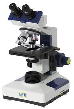
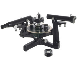
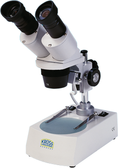
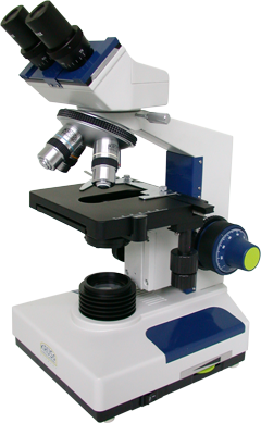
A wide range of Microscopes for Biology, Laboratory and Industry
Krüss offers Microscopes for biological examinations andStereo Microscopes for applications in laboratories, industry and education – ranging from low-cost compact models for general usage, to professional expandable devices for more challenging applications. There is also a metallurgical version – ideal for use in the identification and analysis of all types of metal, including steel and alloys.
Accessories include adapters for video cameras, photo cameras, image processing and connecting cables.
Further information on Krüss Microscopes for the Biology Laboratory can be found here, and information on Stereo Microscopes for quality control and education can be found here.
For prices and availability information on any Krüss Laboratory Microscopes, please contact your nearest distributor, who will be delighted to help.
![]() For full technical specifications, please download the brochure here.
For full technical specifications, please download the brochure here.

KRÜSS microscopes in News and Blog
- Needle-eye art made visible through Krüss microscope
- Microscopes from A.KRÜSS in action in marine biology school projects
- Krüss microscopes at the Hamburg Academy for naturopathy
How have microscope lenses developed?
In any optical instrument like a light microscope, the quality of the image depends most of all on the quality of the optics. Early microscopes used simple single lenses, ground by pioneering glass workers in Europe. These revealed a previously hidden world of tiny detail but, as the ambition of microscopists grew, presented a fundamental limitation. Lenses function by refracting light and, of course, different wavelengths of light refract differently. This phenomenon gives rise to chromatic aberration, the distortion of an image because the different colours of light are brought into focus at different distances from the lens element. An image with chromatic aberration shows rainbow fringes of colour around the subject, limiting the detail that can be resolved.
Among the great scientists who were defeated by this problem was Isaac Newton, who even declared that it was impossible to correct for chromatic aberration. However it was discovered that a doublet – two lens elements made out of different optical materials – could be made to focus two colours on the same plane. This type of lens is today known as the achromatic lens, and is found on even the most basic of optical instruments.
The development of the achromatic lens led to the apochromatic element, which is designed to bring three wavelengths into focus simultaneously. Typically these wavelengths are red, green, and blue, to build a better colour image than the simpler achromatic lens.
The ultimate development in optical microscopy today is the planachromatic lens, sometimes called the superachromatic lens. This combines chromatic corrections at four wavelengths with corrections for spherical aberration. The fourth wavelength is generally in the infra-red region to obviate the need to refocus when conducting multispectral imaging.


